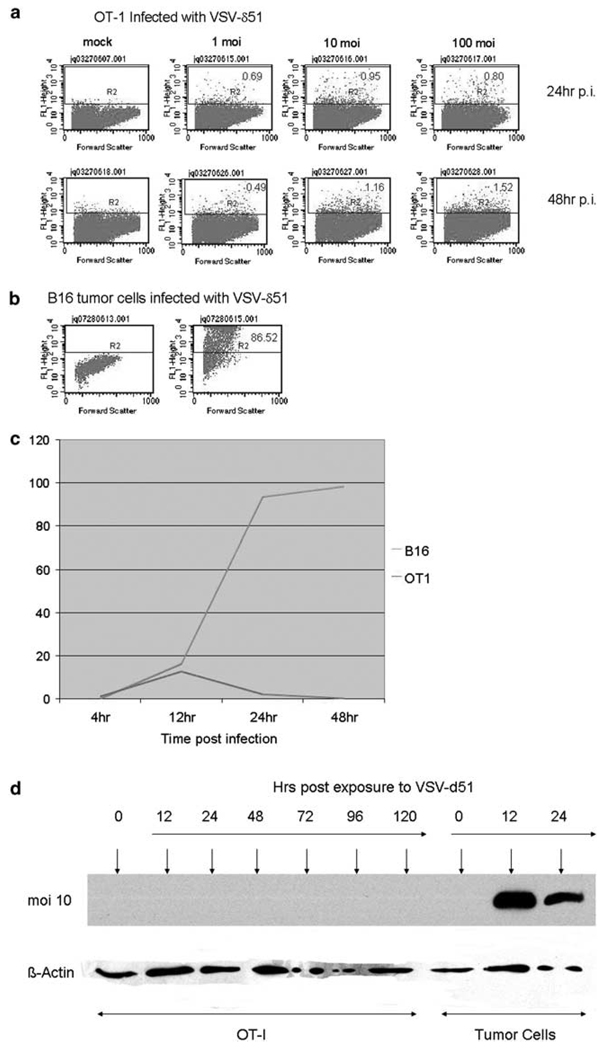
Figure 1.
Vesicular stomatitis virus (VSV)-δ51 infects OT-I T cells at very low efficiencies. (a, b) Three-day-activated OT-I cells, or (b) B16 tumor cells were exposed to cell-free stocks of VSV-δ51, which encodes a green fluorescent protein (GFP) reporter gene, at MOI of 1, 10 or 100 as shown. Cells were analyzed 24 or 48 h later for GFP expression. (c) Three-day-activated OT-I, or B16 tumor, cells were exposed to cell-free stocks of VSV-δ51 at an MOI of 1. Infected cultures were analyzed at time points shown using flow cytometry for viral-derived GFP expression. The percentage of cells in each culture that were GFP+ are plotted with time. (d) Three-day activated OT-I, or tumor, cells were exposed to cell-free stocks of VSV-δ51 at an MOI of 1. Cell lysates were prepared at the times indicated and probed for expression of VSV-G or β-actin using western blotting.

