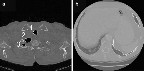
Fig. 1.
2D-segmentation inaccuracies from.23a Top of the thorax: the two lung sections labelled 2 and 3 might be wrongly classified as belonging to different organs, because they are not 2D-connected. b This slice is taken from the low part of the thorax, and shows also the liver and the spleen: the left-lung sections (right part of the image, owing to radiological convention) appear fragmented.

