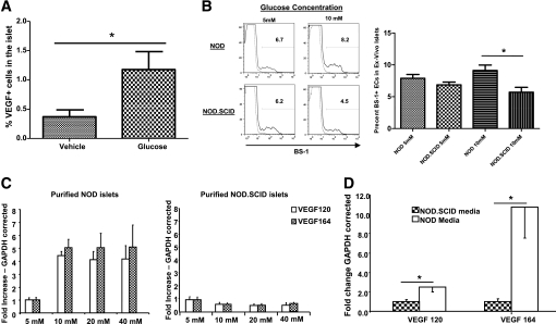
FIG. 4.
High glucose levels stimulate VEGF expression under the conditions of islet inflammation. A: Histomorphic analysis of islets from the pancreatic section of vehicle- or glucose-treated anti-CD3–treated NOD mice (P < 0.02). B: Purified islets from 8- to 10-week-old NOD or NOD.SCID mice were incubated in either 5 or 10 mmol/L glucose for 7 days. After incubation, islets were dispersed and endothelial cell frequency was analyzed using fluorescence-activated cell sorter of BS-1+ cells as described in research design and methods. Left panel: A representative histogram depicting a sample of two NOD or NOD.SCID islet cultures stained for BS-1. Right panel: Summary of the percentage of BS-1+ cells after culture in 5 or 10 mmol/L glucose. n = 6 per group; *P < 0.013. C: Purified islets from 8- to 10-week-old NOD (left) or NOD.SCID (right) mice as in B were incubated with increasing concentrations of glucose. Quantitative RT-PCR was done for VEGF-120 and VEGF-164 isoforms in the islets. D: Supernatants from NOD or NOD.SCID islets incubated at 10 mmol/L glucose for 72 h were added to freshly purified NOD.SCID islets. Quantitative RT-PCR was done for VEGF-120 and VEGF-164 isoforms in the islets (*P < 0.05). n ≥ 3 per group.

