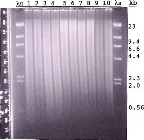
Figure 5:
Agarose gel (Fall 2009) of Canton S (CS) and dusky73 (dy73) genomic DNA digested with different restriction enzymes. Approximately 4 μg of CS DNA (lanes 1, 3, 5, 7, and 9) and 4 μg of dy73 DNA (lanes 2, 4, 6, 8, and 10) were digested with EcoRI (lanes 1 and 2), PstI (lanes 3 and 4), XhoI (lanes 5 and 6), BamHI (lanes 7 and 8), or BamHI and SalI (double digest; lanes 9 and 10) and size separated overnight through 0.8% agarose (1 × TAE). Standard marker on the outside lanes is HindIII-digested lambda DNA (λH; 1.25 μg). The gel was stained with EtBr, destained in 1 × TAE, and visualized and photographed (Polaroid, 667 film) on an ultraviolet transilluminator. The Polaroid print was digitally scanned and enhanced by Photoshop. Before being blotted, the gel was cut in two (between lanes 4 and 5), and each part was given to two of the six student groups for blotting. The Southern hybridizations of these blots are shown together in Figure 6.

