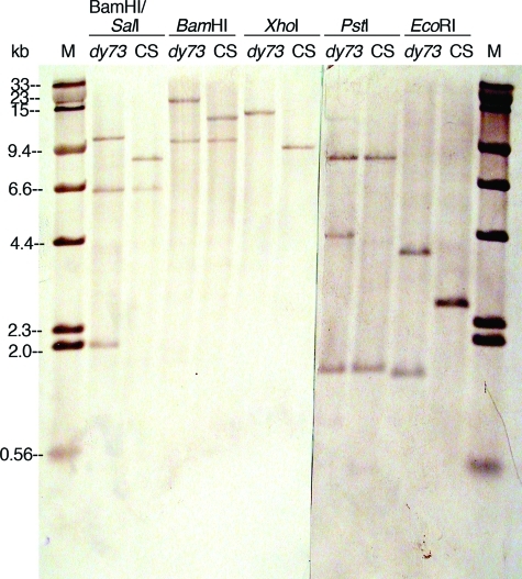
Figure 6:
Southern hybridizations showing RFLPs between restriction enzyme–digested Canton S (CS) and dusky73 (dy73) genomic DNAs. The gel shown in Figure 5 was sliced in two and blotted onto nylon membranes, hybridized with biotinylated plasmids containing a 2.6-kb EcoRI-digested fragment of the CS genome, and detected with streptavidin–alkaline phosphatase conjugate, BCIP, and NBT. The blots (mirror images of the gels due to the blotting procedure) were paired up and digitally photographed (Nikon Coolpix995 camera). This photograph (which was posted online for student use) was enhanced (autolevels, contrast, brightness, and annotation) with Photoshop. The standard marker (M) is a mixture of biotinylated HindIII-digested lambda DNA and SalI-digested lambda DNA that had been size separated and blotted with the 1.25-μg HindIII-digested lambda DNA visible on the EtBr-stained gel (Figure 5).

