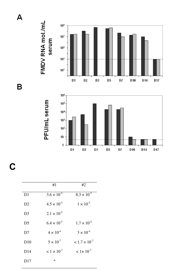
Figure 3.
FMVD replication in serum. Two pigs were bled at each time point. FMDV has been quantified by quantitative RT-PCR and titration in BHK-21 cells. A. It is represented the number of FMDV RNA molecules/mL of serum. The dotted lined indicates the detection limit of the technique (103 FMDV RNA molecules). B. Bar graph indicated the number of PFU/mL of serum quantitated by plaque assay on BHK-21 cells (see Materials and methods). Each bar represents one animal. At 3 dpi one animal of the group was found dead, reason why we did not collect serum from that animal. The dotted line indicates the detection limit of the technique (5 PFU). Each bar represents one animal. C. The specific infectivity is indicated per each animal. It is expressed as the number of PFU per viral RNA molecule.

