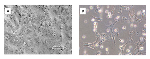
Figure 5.
Appearance of MDCK and Mv1 Lu cells 48 hr after infection of the cells at a high MOI with a pandemic H1N1 2009 virus. Enlarged nuclei and cellular granulation but no cellular detachment are evident in the MDCK cells (panel A), whereas most of the Mv1 Lu cells have detached or are showing advanced CPE (panel B).

