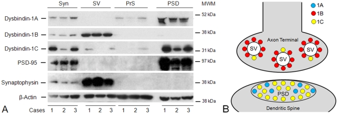
Figure 5. Subsynaptic localization of dysbindin-1 isoforms.
A: Western blotting results on synaptosomal (Syn), synaptic vesicle (SV), presynaptic membrane (PrS), and postsynaptic density (PSD) fractions of the normal HF from three control cases (1–3) in this study. Successful synaptic fractionation was confirmed with synaptophysin as a marker for synaptic vesicles and PSD-95 as a marker for the PSD. No selective molecular marker is available for the PrS fraction, including syntaxin-1 since this proves to be ubiquitously distributed on subcellular membranes. β-actin served as the loading control. The blots were probed with PA3111 (1∶1000) for dysbindin-1A and -1C and with UPenn 331 (1∶5000) for dysbindin-1B. In synaptosomes, dysbindin-1A and -1C were found to be predominantly PSD proteins, while dysbindin-1B was found to be predominantly a synaptic vesicle-associated protein. MW = molecular weight marker position. B: Diagram of pre- and/or post-synaptic location of dysbindin-1 isoforms consistent with the Western blotting results just shown and with dysbindin-1 immunohistochemical findings at the electron microscopic level by Talbot et al. [43].

