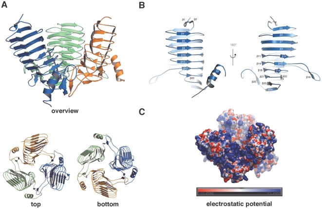
Figure 2. Crystal structure of NeuO.
(A) Overview about the three-dimensional structure of NeuO with each monomer displayed in a different color in cartoon mode; top and bottom views are given. Monomers are tilted by almost 45° with respect to each other. (B) A single monomer of NeuO is displayed from both sides; the annotation for every secondary structure element is indicated. (C) Representation of the electrostatic surface potential of the NeuO trimer, displayed from −15 to 15 kbT, calculated using the program APBS. [44]. Most of the surface is positively charged (blue) suited to bind to the poly-anionic acceptor substrate polySia.

