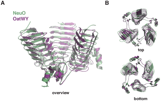
Figure 4. Structural comparison between NeuO and OatWY.
A least-squares superimposition of NeuO (green) (PDB-ID: 3JQY) and OatWY (magenta) (PDB-ID: 2WLD) reveals the high overall similarity between both molecules. Irrespective of their different substrate specificities the monomers of both proteins can be superimposed with an r.m.s.d. of 0.9 Å. Globally both proteins share a high inclination between the chains, probably as an adaptation to their unusually long substrates. The least-squares superimposition was calculated using the program LSQMAN [45].

