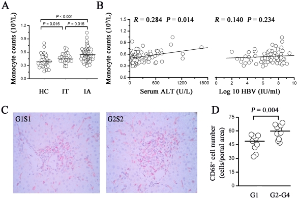
Figure 1. Increased monocytes in peripheral blood and liver positively correlate with liver injury in IA patients.
(A) Absolute counts of the peripheral blood monocytes from IT (n = 32), IA (n = 78) patients and healthy controls (n = 38). Each dot represents the value from one individual. P values are shown. (B) The absolute numbers of peripheral blood monocytes was significantly correlated with serum ALT levels but not with HBV DNA. The solid line represents the linear growth trend and r, the correlative coefficient. P values are shown. (C) Representative immunohistochemical staining of CD68 in specimens from IA patients. An IA patient with HAI G2S2 scores has higher CD68+ cell density in liver, compared to that of a patient with HAI G1S1 scores. (D) Numbers of CD68+ cells in portal area are shown in IA patients with various degrees of liver HAI scores. Horizontal bars represent the median CD68+ cells. ALT, alanine aminotransferase; HAI, histological activity index; IT, Immune tolerant carriers; IA, immune activated patients.

