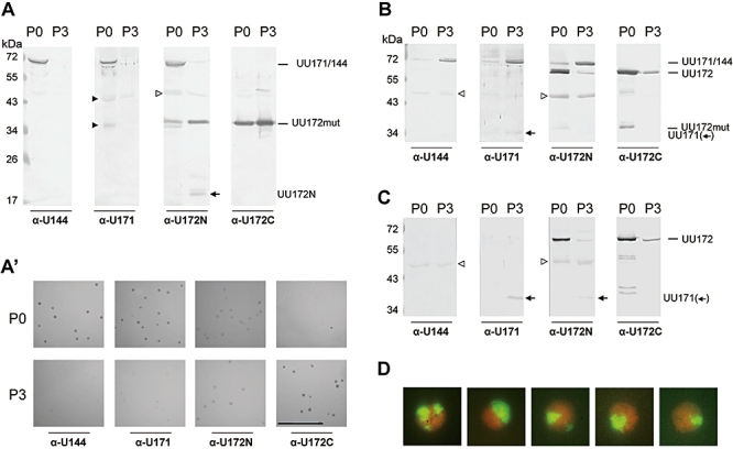
Fig. 5.

Phase variation of clonal variants of U. parvum serotype 3 before and after exposure to monospecific Pabs.
A–C. Western blot analyses of: (A) M14-K16 before (P0) and after (P3) serial exposure to Pab α-U144, (B) DR1-K1 before and after serial exposure to Pab α-U172C, and (C) 27815T-K5 before and after exposure to Pab α-U172C. Proteins were detected with monospecific Pabs (see Table S3) indicated below the blots. Black triangles: cross-reaction with the UU376 protein. Open triangles: background bands, which were already detectable with pre-immune sera.
A′. Colony immunoblots of M14-K16 before and after serial exposure to Pab α-UU144. Cells were plated before antibody treatment (P0) and after the third passage (P3) after antibody treatment. Scale bar: 1 mm. For immunostaining, monospecific Pabs (see Table S3) indicated below the blots were used.
D. Colony immunoblots showing sectored colonies of subclonal populations of M14-K16, which had been exposed to Pab α-U144. Double immunofluorescence overlay: green (bright) sectors, UU171/144 expression and red (dark) sectors, UU172 expression.
