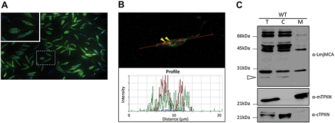
Fig. 1.

LmjMCA intracellular localization.
A. Endogenous LmjMCA localization was analysed by immunofluorescence using an anti-LmjMCA antibody (green). Nuclei and kinetoplasts were stained with DAPI (blue).
B. Confocal microscopy of LmjMCA-overexpressing parasites. Mitochondrion was stained with Mitotracker (red) and a signal/distance profile was generated in order to confirm the colocalization.
C. L. major parasites subcellular fractionation was obtained by digitonin treatment. T (total) corresponds to total proteins, C (cytoplasm) corresponds to 100 µM digitonin and M (mitochondria) corresponds to 500 µM digitonin. Proteins were immunoblotted using RE53.
Anti-mTPXN (mitochondrial tryparedoxin peroxydase) anti-cTPXN (cytoplasmic tryparedoxin peroxydase) were used as fractionation controls.
