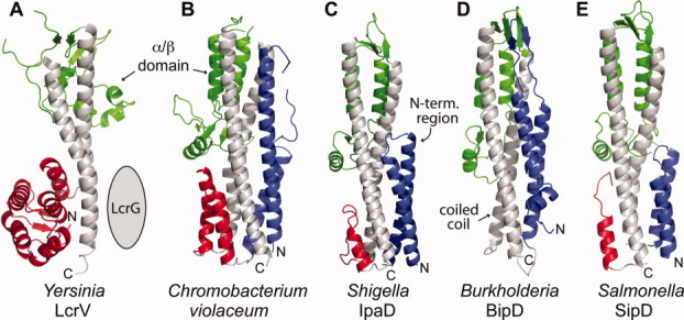
Figure 5.

Comparison of five current crystal structures of T3SS tip proteins: (A) Yersinia LcrV,18 (B) a SipD homolog from Chromobacterium violaceum, (C) Shigella IpaD,4 (D) Burkholderia BipD,4,5 and (E) Salmonella SipD. The central coiled coil (gray) and mixed α/β domain (green) are common structural features of T3SS tip proteins. The N-terminal region in (A) forms a globular domain of α-helices and β-strands (red), whereas in (B–E), the N-terminal region forms α-helical hairpins (blue) followed by short α-helices (red) in (B, D, and E).
