
Figure 7.
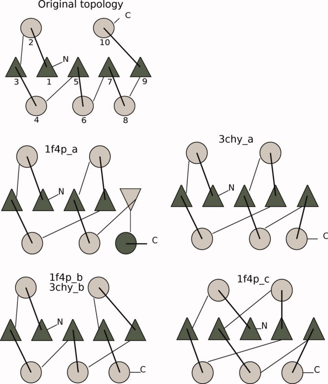
Rossmann fold topology, original, and the four different topologies (two different protein probes). Triangles represent β-strands, and circles represent α-helices. The colors designate the direction of the SSEs, dark gray starting at the back of the page coming at you, and light gray starting in front and traveling back through the page (N-C terminal). The SSEs of the original fold are enumerated in the N-C terminal direction. [Color figure can be viewed in the online issue, which is available at wileyonlinelibrary.com.]