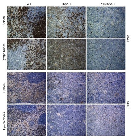
Figure 3.
Immunohistochemical staining of lymphomas involving the spleen and lymph nodes in iMycEµ and K13/iMycEµ. Tissue sections were prepared from spleens and lymph nodes of WT and tumor (T)-bearing iMycEµ and K13/iMycEµ mice and stained with B220 and CD3 antibodies as described in the materials and methods. The lymphoma-infiltrated spleens and lymph nodes from K13/iMycEµ mice appear negative for B220 and CD3. The scale shown is ×200.

