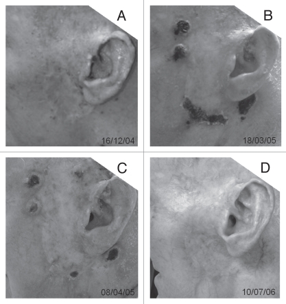
Figure 2.
Photos of lesion of one patient with stage III C melanoma around the left ear treated by in situ photoimmunotherapy (ISPI). (A) Before ISPI, showing extensive acute radiation damage and numerous small black metastases. (B) Immediately after the first laser treatment in the second cycle of ISPI, bullae can be seen after laser treatment. (C) Immediately after the second laser treatment in the second cycle of ISPI. (D) All treatment-related ulcers have healed and the subject is free of clinically and radiologically detectable tumors after completion of three cycles of ISPI.

