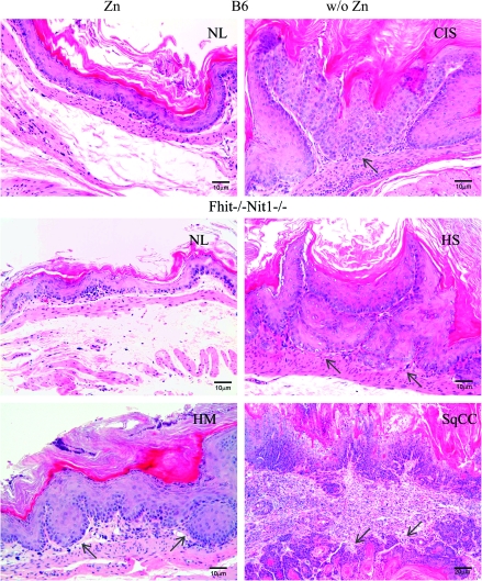
Fig. 2.
Histopathology of representative forestomach tissues of unsupplemented and Zn-supplemented mice. In the left panels from mice that received Zn supplementation immediately after final carcinogen treatment are photomicrographs of hematoxylin- and eosin-stained forestomach tissues: normal mouse forestomach (NL) tissue sections with two to three layers keratinizing stratified squamous epithelium without cytological and nuclear atypia or architectural dysplasia (left, top two panels) and mild hyperplasia (HM, lower left panel) in which the squamous epithelium is up to 10 layers thick and focal nuclear atypia may be present, indicated by arrow. In the right panels from mice that did not receive Zn supplementation are photomicrographs of hematoxylin- and eosin-stained forestomach tissues: CIS (carcinoma confined to epithelium with intact basement membrane, top right panel), indicated by arrow; severe hyperplasia (HS, squamous epithelium >10 layers thick with or without nuclear atypia and papillomatous change, middle right panel), indicated by arrows; squamous cell carcinoma (SqCC, right bottom panel) with invasion through the basement membrane; arrows in SqCC denote well-differentiated invasive SqCC with keratin pearls. The tumor cells show loss of maturation and loss of nuclear polarity. Nuclei are enlarged with high nuclear/cytoplasmic ratio and denser hyperchromasia. Mitotic activity is frequently present and atypical keratinizing squamous epithelium is also seen.

