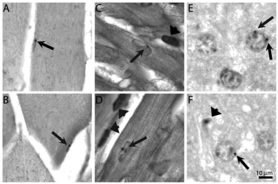
Figure 1.
Bone marrow-derived cells in quiescent tissue. Brightfield images of tissue sections after in situ hybridization with digoxygenin labeled pBR322 probe to detect bone marrow-derived cells in chimeric mice. A & B) Liver section of young (A) and diabetic (B) mouse methyl green stained to delineate nuclei. Arrows point to small black dots indicating hybridization to pBR322 probe to hepatocyte nuclei. Arrowhead in (B) shows labeled cell, possibly a Kupffer cell. C & D) Desmin labeled sections of skeletal muscle with labeled nuclei (arrows) in young mice. E & F) Desmin labeled sections of myocardium with labeled nuclei (arrows) in an old (E) and young (F) heart. Bar = 10 μm.

