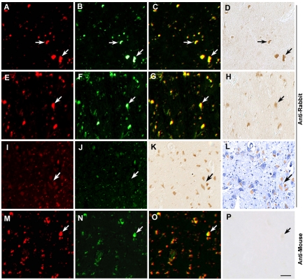
Figure 2.
Autofluorescent characterization and immunohistochemical localization of the cross-immunoreactivity with anti-rabbit secondary antibody in aged human brain tissues. Bright autofluorescence (arrows) in hippocampal regions (A-C), and parietal cortex (E-G). Lipofuscin autofluorescence (arrows) is detected in both Cy3 and FITC filter channels (in a yellow color on the merged images), and produces cross-immunoreactivity with anti-rabbit antibody in the absence of any primary antibody (D and H). In the aged human cortices, high autofluorescence is associated with moderate cross-immunoreactivity (A-H), whereas diffuse and weak autofluorescence is associated with high cross-immunoreactivity (I-K). Counterstaining with hematoxylin (L) demonstrates that nonspecific immunoreactivity is localized to the cytoplasm. No cross-immunoreactivity is detected with anti-mouse antibody (M-P). Scale Bar 30 μm.

