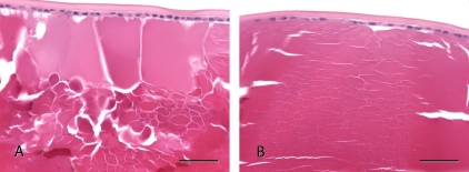
Figure 4.
Histologic appearance of the anterior lens of mice. (A) A focal, minimal, unilateral area of liquefaction and globule formation in the anterior cortex of the lens of an ASIC1a+/+ mouse at 23 weeks. (B) Slight swelling of lens fibers in the anterior cortex of the lens of ASIC1a-/- mouse at 27 weeks of age. H&E stain. Bar = 50 μm.

