
Fig. 1.
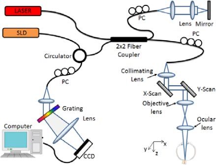
Schematic of the OMAG system used for collecting the spectral interferogram data sets to perform 3D angiogram and quantitative blood flow measurement of the rat retina in vivo. CCD: the charge coupled device, PC: polarization controller. Raster-scanning both the X and Y-scanner, we can collect a 3D volume data set. By keeping one of the scanners static, repeated B-scan (i.e., M-B scan) could be achieved.