
Fig. 2.
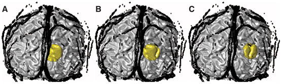
3D coronal view of the anatomical head model including the extra-cerebral vasculature (black), gray matter (GM) and the region of activation (gold). The distance between the midline and the center of regions A, B and C were 10, 20 and 30 mm, respectively.