Fig. 1.

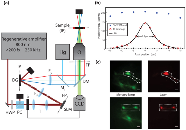
(a) Microscopy Setup. HWP: half-wave plate, PC: polarizer cube, S: camera-controlled shutter, T: telescope comprised of two achromatic lenses (f = −75 mm and +125 mm), SLM: spatial light modulator, FP: Fourier plane, FF: Fourier achromatic lens (f = 300 mm), DG: diffractive grating, IP: image plane, FG: achromatic lens for collimating the diffracted beam (f = 300 mm), Hg: mercury arc lamp, M1: Flip mirror, DM: excitation-detection dichroic filters, O: microscope objective, CCD: EMCCD camera. (b) Fluorescence intensity of a thin layer of fluorescein scanned through the focal plane with and without TF by replacing the DG by a mirror. (c) Fluorescence of live cultured cells loaded with Fluo-4 AM calcium indicator. (Green) Wide-field mercury arc lamp excitation (λexc = 450 nm, λem = 525 nm longpass), (Dashed line) targeted illumination, (Red) TF patterned illumination (λexc = 800 nm, λem = 535/40 nm). All scale bars are 20 µm.
