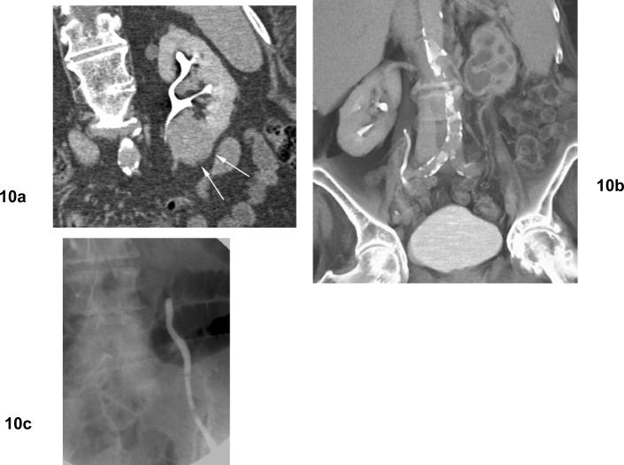
Fig 10. Ureteral stricture and hydronephrosis after cryoablation for clear cell type renal cell carcinoma.
(a) Excretory phase coronal CT shows mass in the lower pole of the left kidney adjacent to the ureteropelvic junction. (b) Excretory phase coronal CT shows left hydronephrosis developed after cryoablation. (c) Retrograde ureterography shows obstruction of the left proximal ureter at the level of the ureteropelvic junction.

