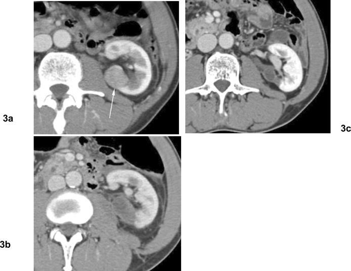
Fig 3. Involution of treated tumor after cryoablation. Papillary type renal cell carcinoma.
(a) Axial contrast enhanced CT before cryoablation shows solid mass in the left kidney posteriorly (arrow). Biopsy showed papillary type renal cell carcinoma. (b) Venous phase contrast enhanced CT 2 months after cryoablation shows heterogeneous hypodensity in ablation zone without contrast enhancement. (c) Venous phase contrast enhanced CT 10 months after cryoablation shows involution of treated tumor without contrast enhancement.

