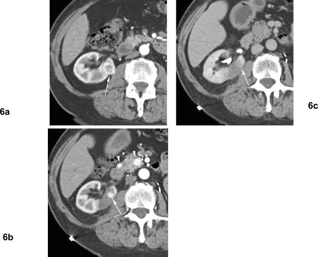
Fig 6. Residual unablated tumor after cryoablation. Clear cell type renal cell carcinoma. The patient is status post left nephrectomy for renal cell carcinoma.
(a) Arterial phase contrast enhanced CT before cryoablation shows heterogeneously enhancing mass (arrow) in the right kidney medially. (b) Arterial phase contrast enhanced CT 1 month after cryoablation shows nodular contrast enhancement at the periphery of ablation zone (arrow) indicating residual unablated tumor. Note enhancing small masses in the pancreas (arrowheads) indicating metastatic foci. (c) Excretory phase contrast enhanced CT 1 months after cryoablation shows washout of contrast material from the nodular enhancement, and unablated tumor is seen as subtle hypodense area relative to normal renal parenchyma (arrow).

