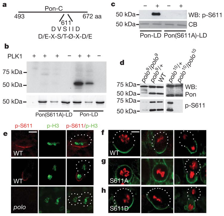
Figure 1. Polo phosphorylates Pon and this phosphorylation event regulates the asymmetric localization of Pon.
a, A diagram of the putative Polo phosphorylation site (S611) in Pon-LD, which matches the consensus Polo phosphorylation site (D/E-X-S/T-Ø-X-D/E, where Ø is a hydrophobic amino acid and X is any amino acid). b, In vitro kinase assays. Upper panel, autoradiography; lower panel, Coomassie blue (CB) staining of the same gel. c, Western blot (WB) analysis testing the specificity of the p-S611 antibody. d, Western blot analysis showing loss of p-S611-positive Pon in polo mutants. Note that, in addition to the 72 kDa full-length Pon protein, both the general Pon antibody and the p-S611 antibody recognized a ~50 kDa band—a putative proteolytic product. The level of the 50 kDa band seems to be dependent on Polo activity. e, Staining of endogenous Pon in wild-type (WT, upper and middle panels) and polo mutant (lower panels) cells using the p-S611 antibody. p-H3, p-histone H3. f–h, Localization of GFP–Pon-LD (WT) (f), GFP–Pon(S611A)-LD (g) or GFP–Pon(S611D)-LD (h) in embryos at stages 9–12. Neuroblasts at different cell cycle stages are shown. GFP–Pon-LD proteins were detected by immunostaining with anti-myc antibody (green). Anti-p-histone H3 staining (red) was used to indicate mitotic stages. Scale bars, 5 μm.

