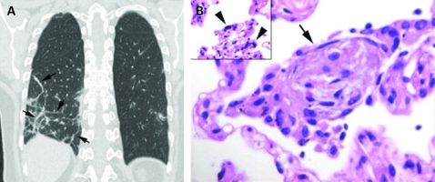
Figure 1.
(A) Coronal reformation computed tomography image showing organising pneumonia in a perilobular distribution (arrows). (B) High power photomicrograph shows a polypoid plug of fibroblastic tissue within an alveolar space (arrow). Note alveolar cell showing viral cytopathic changes (arrowheads).

