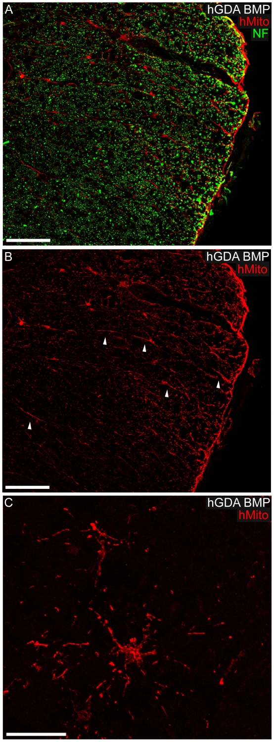
Figure 4. Migration and morphology of hGDAsBMP within DLF white matter.
(A, B) High power images showing hMito+ hGDAsBMP in the process of migrating and accumulating at the pial surface within lateral funniculus white matter. Note the elongated radially orientated processes displayed by some hMito+ hGDAsBMP within white matter (B: arrowheads), a glial morphology indicative of tangential migration of these cells towards the adjacent pial surface (hMito: red; NF+ axons: green). (C) hGDAsBMP displaying typical astrocytic “stellate” arrangements of their processes within white matter immediately ventral to the injury site. Survival = 5 weeks post injury/transplantation. Scale bars: A, B = 100 µm; C = 20 µm.

