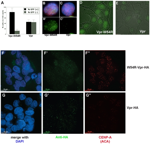
Figure 1. Transient transfection to induce Vpr expression destabilizes both endogenous and GFP-CENP-A.
A. Manual quantitation of GFP-CENP-A cells after transfection with control Vpr mutant W54R or wild-type Vpr. B–C . Example images of cells transfected with control Vpr-W54R-HA (B-B
 ) or wild-type Vpr-HA (C-C
 ) detected with anti-HA in red, DNA in blue, and GFP-CENP-A in green. GFP-CENP-A channel is also shown alone (B
 and C
and C ). D–E. Example fields of live GFP-CENP-A cells transfected with Vpr-W54R (D) or wild-type Vpr (E). F–G
 . Example fields of fixed HeLa cells after transfection with Vpr-W54R (F-F
 ) or wild-type Vpr (G-G
 ). Vpr is detected with anti-HA in green. Endogenous CENP-A is detected with anti-centromere autoantisera (ACA) in red.

