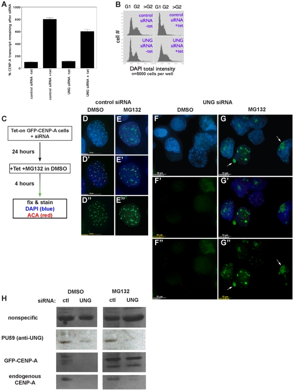
Figure 3. Loss of newly-made GFP-CENP-A after UNG-directed siRNA treatment is due to mislocalization and destabilization, not cell-cycle arrest or lack of synthesis.
A. Quantitative real-time RT-PCR was used to detect levels of CENP-A transcript (endogenous and GFP-CENP-A combined) with and without UNG-directed siRNA or tetracycline. Percent CENP-A transcript levels were normalized against the sample with control siRNA without tetracycline. B. Cell cycle analysis was performed using high-content automated image quantitation, with and without UNG-directed siRNA or tetracycline. C–G . GFP-CENP-A cells were transfected with UNG-directed siRNA were treated with MG132 for 4 hours to prevent protein turnover. A scheme is shown in part C, example DMSO-treated control cell (D-D
 ), example MG132-treated control cell (E-E
 ). Example fields are shown for UNG-siRNA transfection with DMSO (F-F
 ) or MG132 (G-G
 ). Endogenous CENP-A was detected with human autoantisera (ACA) in red. H. Western blot analysis of protein levels demonstrates that GFP-CENP-A is selectively stabilized by MG132 treatment after UNG reduction by siRNA, while endogenous CENP-A protein levels remain low.

