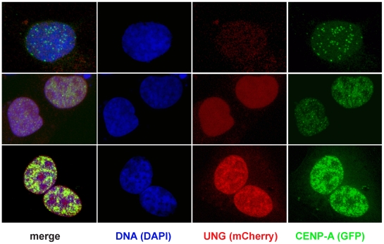
Figure 11. Overexpressing UNG-mCherry is sufficient to rapidly induce excess accumulation of GFP-CENP-A.
Example images of 143b cells transiently co-transfected with mCherry-UNG (red) and GFP-CENP-A (green), fixed and stained with DAPI to detect DNA (blue). In cells with no detectable UNG, GFP-CENP-A localized to centromeres (top row). In cells with low levels of mCherry-UNG, GFP-CENP-A was distributed throughout the nucleus (middle row). In cells with high levels of mCherry-UNG, GFP-CENP-A accumulated to extremely high levels (bottom row).

