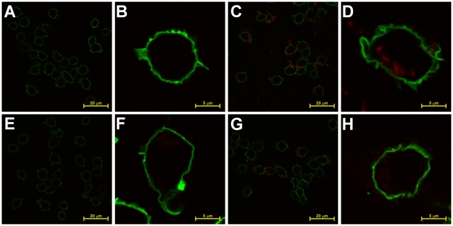
Figure 8. Detection of intracellular and extracellular bacteria by microscopy of single copy expression of PcysTomato.
(A–B) Y. pestis KIM D27, (C–D) pRsaI-2.1, (E–F) KIM D27-1013 (dapAX attTn7::dapAX PcysDsRed), or (G–H) KIM D27-1014 (attTn7::dapAX cys-Tomato) were grown overnight in HIB at 26°C, diluted 1∶15 in HIB and grown for 2 hours, and then used to infect biotinylated RAW 264.7 macrophage-like cells at an MOI of 10 for 30 minutes. Cells were then fixed and stained with streptavidin conjugated to AlexaFluor 488 to indentify the host cell membrane. Samples were analyzed by laser scanning confocal microscopy.

