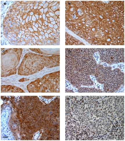
Figure 1. Immunoexpression of pEGFR, HER3, HER4, pAkt, Akt1 and PTEN proteins in PSCC.
Strong immunoexpression of pEGFR (A), HER3 (B) and HER4 (C). pAkt (D) and Akt1 (E) show both nuclear and cytoplasmic staining. PTEN expression is restricted to nuclei only and reduced staining was often found in cancer cells (F). Scale bar: 50 µm.

