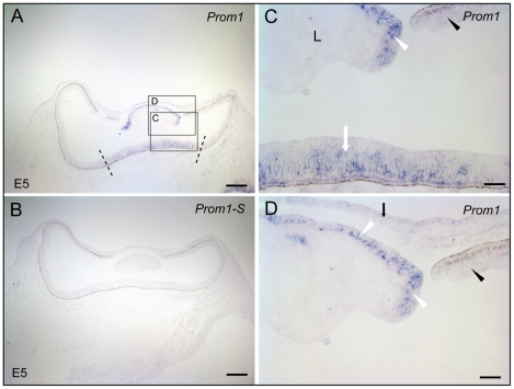
Figure 4. Localization of prominin-1 in the eye of chick at the embryonic day 5.
(A–D) Cryosections of chick embryos (E5) were processed for non-radioactive ISH using either antisense (A, C, D; Prom1) or sense (B; Prom-1S) DIG-labelled prominin-1 probe. The boxed areas in panel A are shown at higher magnification in panels C and D. Dashed lines delimit the central retinal primordium and white arrow indicates prominin-1 expression. Black arrowhead and arrow point to the peripheral retinal primordium and corneal epithelium, respectively, being weakly positive for prominin-1. White arrowheads indicate the prominin-1–positive anterior lens (L) epithelium. Scale bars, A and B, 250 µm; C and D, 50 µm.

