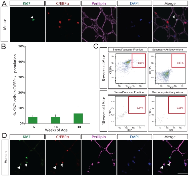
Figure 2. Cellular replication in adipose tissue assayed with the cell cycle protein Ki67.
A. In mouse adipose tissue, Ki67 (green) overlaps with C/EBPα (red) and DAPI (blue) in perilipin-positive (purple) adipose tissue in situ. White arrow indicates triple positive cell. Isotype antibody control shown in right most panel. Scale bars 50 µm. B. The percentage of Ki67-positive C/EBPα-expressing cells was determined in 6 (n = 6), 14 (n = 9), and 30-week old (n = 3) mice. Throughout adult life, 4.8% of cells in adipose tissue are Ki67 and C/EBPα-positive at any time. C. FACS plots of the stromal/vascular nuclei from dissociated fat tissue of wild-type mice, stained for Ki67 and C/EBPα, as well as appropriate secondary antibody controls. The total percentage of Ki67-positive C/EBPα-positive nuclei in the stromal/vascular fat sample (above secondary alone background for all antibodies) was 3.85%; of this 0.06% represented C/EBPα-high Ki67-positive events, and 3.79% represented C/EBPα-low Ki67-positive events. Top panels represent 8-week-old mice, bottom panels represent 10-week-old mice. D. In human adipose tissue, Ki67 (green) overlaps with C/EBPα (red) and DAPI (blue) in perilipin-positive (purple) adipose tissue in situ. White arrow indicates triple positive cell. Scale bars 50 µm.

