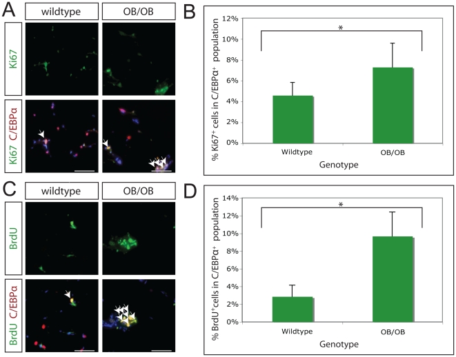
Figure 4. Increased adipose tissue replication in OB/OB mice.
A. The rate of adipose tissue replication was assayed by co-staining for the cell cycle protein Ki67 (green) and the preadipocyte/adipocyte marker C/EBPα (red) in 14-week-old wildtype and OB/OB mice. Representative images of wildtype and OB/OB adipose tissue are shown. White arrows indicate cells co-staining for Ki67 and C/EBPα. DAPI stained nuclei are blue. Magnification, 200x; scale bars 100 µm. B. The percentage of C/EBPα-expressing cells in the growth cycle was quantitated for 14-week old C57/BL6 wildtype (n = 9) and OB/OB (n = 6) mice. There is a statistically significant increase in the rate of preadipocyte replication (p<0.05) from 4.6% to 7.3%, in wildtype and OB/OB mice, respectively. C. The rate of adipose tissue replication was assayed by co-staining for BrdU incorporation (green) and the preadipocyte/adipocyte marker C/EBPα (red) in 14-week-old wildtype and OB/OB mice. Representative images of wildtype and OB/OB adipose tissue are shown. White arrows indicate cells co-staining for BrdU and C/EBPα. DAPI stained nuclei are blue. Magnification, 200x; scale bars 100 µm. D. The percentage of C/EBPα-expressing cells having incorporated BrdU following 5 days of BrdU injection was quantitated for 14-week old C57/BL6 wildtype (n = 5) and OB/OB (n = 6) mice. There is a statistically significant increase in the rate of adipose tissue replication (p<0.05) from 2.9% to 9.7%, in wildtype and OB/OB mice, respectively.

