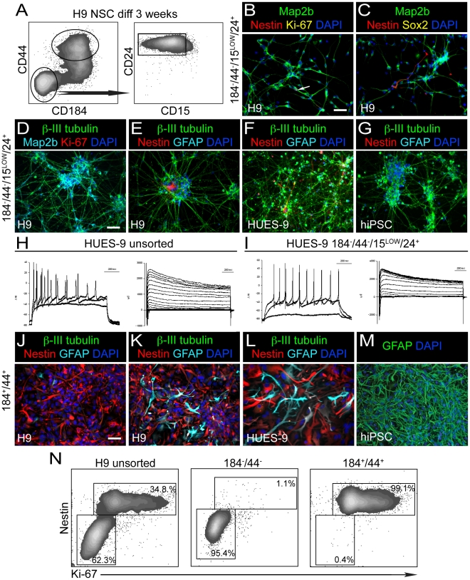
Figure 3. Sorting neurons and glia from cultures of sorted and subsequently expanded and differentiated NSC.
NSC were differentiated for 3 weeks in neuron differentiation medium prior to FACS. (A) Cell sorting strategy of differentiated H9 NSC using CD184−/CD44−/CD15LOW/CD24+ and CD184+/CD44+. Similar populations were isolated from differentiated cultures of HUES-9 NSC and NDC3.1 NSC both derived from SDIA PA6 co-culture. (B) Sorted CD184−/CD44−/CD15LOW/CD24+ cells were cultured in neuron differentiation medium for 2 days post-FACS and subsequently stained with anti-Map2b, anti-Nestin, anti-Ki-67 and DAPI. White arrow indicates the presence of one Ki-67+, Nestin+ cell in this field. (C) Same as B except cells were stained with anti-Sox2 instead of Ki-67. One Nestin+ cell is evident in this field. (D) CD184−/CD44−/CD15LOW/CD24+ sorted cells were cultured in neuron differentiation medium for 7 days post FACS and subsequently stained with anti-β-III tubulin, anti-Map2b, anti-Ki-67 and DAPI. No Ki-67+ cells are observed in this field. (E) Same as D but stained with anti-β-III tubulin, anti-Nestin, anti-GFAP and DAPI. No GFAP+ cells are evident and one Nestin+ cell is evident in this field. (F) Same as E but from HUES-9. (G) Same as E but from hiPSC, NDC3.1. (H) Electrical recordings of patched neurons from a 3-week differentiated HUES-9 NSC culture. All cells exhibited Na+ current (right panel). 7 of 8 cells tested fired action potentials when depolarized with current (left panel). (I) Electrical recordings of sorted neurons from a 3-week differentiated HUES-9 NSC culture. Sorted neurons were cultured for an additional 3 weeks prior to recording. All cells exhibited Na+ current (right panel). 6 of 8 cells fired action potentials when depolarized with current (left panel). (J) CD184+/CD44+ sorted cells derived from H9 were cultured in neuron differentiation medium for 7 days post-FACS and stained with anti-β-III tubulin, anti-Nestin, anti-GFAP and DAPI. (K) Same as H except cells were cultured in astrocyte culture medium for 7 days prior to imaging. (L) Same as K, but from HUES-9. (M) CD184+/CD44+ cells from NDC3.1 were cultured for 6 passages in astrocytes medium and stained for GFAP and DAPI. Scale bar is 50 µm. (N) 2-color intra-cellular FACS analysis of NSC sorted from H9 and then induced to differentiate for 3 weeks prior to sorting cell populations based on different combinations of CD184 and CD44 immunoreactivity. The unsorted population is composed of a Nestin+ population that is highly enriched for mitotic Ki-67+ cells whereas the Nestin−/LOW population is quiescent. Sorting of a CD184−/CD44− population enriches for Nestin−/LOW quiescent cells. Isolation of CD184+/CD44+ and CD184+/CD44− cells enriches for the Nestin+ cycling population. Percentages for these 2 populations are indicated.

