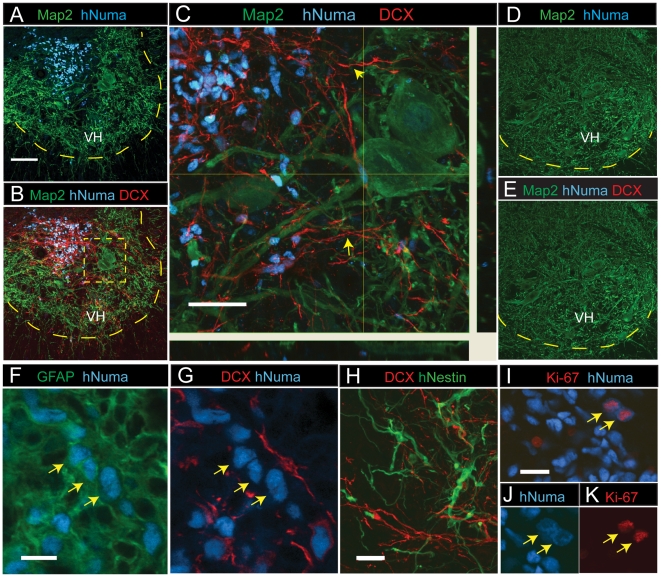
Figure 4. Spinal transplantation of differentiated NSC cultured cells in rats with spinal ischemic injury.
(A) In animals with previous ischemic injury hNUMA+ grafted cells (blue) were identified in the intermediate zone or in the ventral horn (VH) 2 weeks after grafting. Scale bar is 100 µm. (B, C) Numerous hNUMA+ cells were DCX immunoreactive and showed extensive projection of DCX+ processes towards the ventral horn (yellow arrows). Scale bar is 40 µm. Yellow dotted box represents expanded view in C. (D, E) In control animals injected with medium only, no hNUMA or DCX immunoreactivity was identified. (F, G) A subpopulation of grafted hNUMA-positive cells showed colocalization with GFAP but were DCX-negative (yellow arrows). Scale bar is 10 µm. (H) At 2 weeks after grafting hNestin-positive cells were seen in the core of the graft and were DCX negative. Scale bar is 20 µm. (I–K) Proliferating cells were identified by colocalization of Ki-67 and hNUMA immunoreactivity and were primarily seen 2 weeks after grafting. Scale bar is 20 µm.

