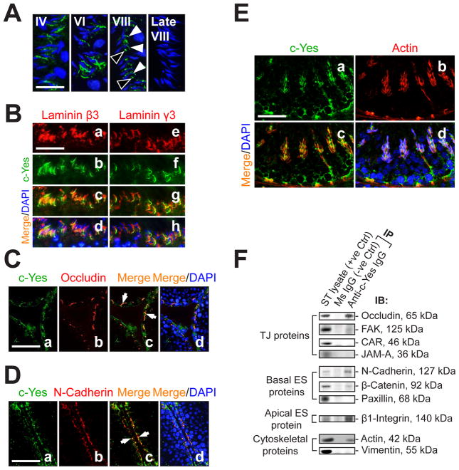
Fig. 2. c-Yes is an integrated component of the BTB and the apical ES as demonstrated by dual- labeled immunofluorescence (IF) analysis and co-immunoprecipitation (Co-IP).
(A) Fluorescence microscopy was used to illustrate stage-specific localization of c-Yes (green, FITC) at the apical ES. Cell nuclei were visualized with DAPI (blue). c-Yes staining was detected at the apical ES surrounding the heads of the developing spermatids throughout the epithelial cycle until shortly before spermiation when it vanished completely (at late stage VIII). It is noted that c-Yes was found to be both at the convex (white arrowheads) and the concave (black arrowheads) sides in early stage VIII, consistent with IHC data shown in Fig. 1. Bar in first panel (also applies to the other three) = 30 μm. (B) Dual-labeled IF analysis confirmed c-Yes occurrence at the apical ES by its co-localization with laminin β3 (a-d) and γ3 (e-h) (red, Cy3), which are known apical ES components expressed exclusively by elongating/elongated spermatids (Yan and Cheng, 2006). Bar in a (also applies to b-h) = 60 μm. (C-D) Presence of c-Yes at the BTB was supported by moderate co-localization (white arrows) with TJ (occludin) (C) and basal ES (N-cadherin) (D) proteins. Bar in a (also applies to b-d) = 100 μm. (E) c-Yes presence at the BTB and the apical ES was further corroborated by its co-localization with F-actin, suggesting it is likely a regulatory component of the structural scaffolding of actin filaments. Bar in a (also applies to b-d) = 40 μm. (F) A study using Co-IP to assess the structural interactions between c-Yes and selected component proteins of the BTB and the apical ES using ~800 μg protein of seminiferous tubule (ST) lysates. Mouse (MS) IgG instead of anti-c-Yes IgG served as a negative control.

