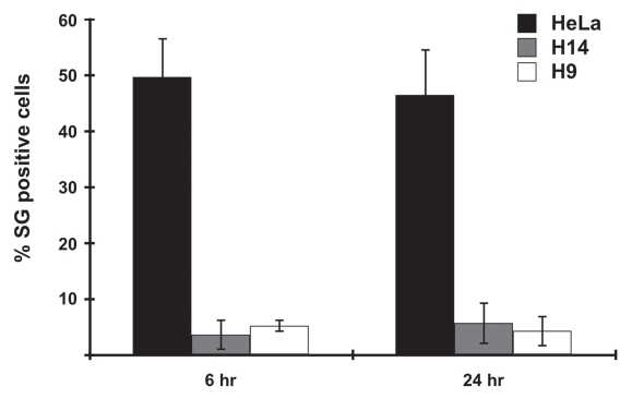
Figure 8.
Fewer visible stress granules (SGs) are formed in hES cells following transfection stress. HeLa, H9 and H14 cells were transfected with 2 µg/mL PIC for 6 hrs and 24 hrs respectively, and then stained with J2 antibody and anti-TIA1. SG-positive cells were counted under 3–6 randomly selected different microscope fields, each containing 100–200 cells. Standard deviation was calculated from all counted fields in three independent experiments.

