Fig. 3.
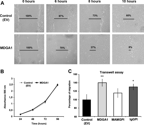
MDGA1 increases cell motility in MDCK cells. a Wound healing assay was performed in MDGA1 and control (EV) cells. Snapshots of the healing were taken at 0, 6, 8 and 10 h, showing MDGA1 cells exhibited an increase in migration (lower lane) compared to control (EV) cells (upper lane). Cell-free area percentages are indicated. b No differences were found in the proliferation rate between MDGA1 and control (EV) cells. c MDGA1 or IgGPI expression increases cell migration rates in Transwell assays compared to control (EV) cells. Values represent mean ± S.D. Significant values are indicated by asterisks

