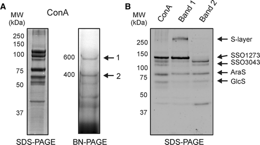
Fig. 4.

Analysis of the sugar binding proteins extracted from membranes derived from S. solfataricus wild-type cells grown on arabinose and purified with ConA lectin affinity purification. ConA fraction analyzed by SDS-PAGE (a, left panel) and Blue-Native PAGE (a, right panel). The sugar binding proteins were present in two complexes with molecular masses of 400 and 600 kDa, respectively. These two complexes were excised, eluted and subsequently run on an SDS-PAGE (b) to show their subunit composition. For comparison the initially isolated Con A fraction is displayed in the first lane. The identity of the proteins was determined by mass spectrometry
