
Fig. 6.
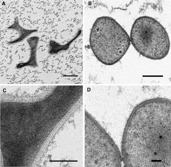
Transmission electron microscopy of ultrathin sections of PBL2025 (a, c) and ΔbasEF (b, d) cells prepared by high-pressure freezing, freeze substitution and embedding in Epon. Sizes of the bars in a and b are 500 nm, c is 200 nm and d is 100 nm