
Figure 1.
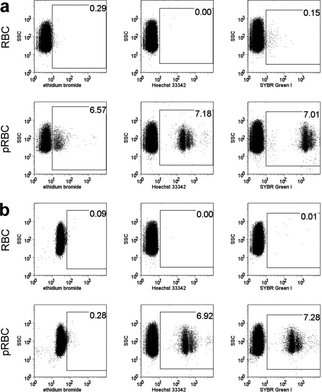
Staining of pRBC with fluorescent DNA-binding dyes. pRBC were detected by flow cytometry after staining with either 10 μg/mL EB, 2 μM Hoechst 33342, or 1:5,000 SYBR Green I. a: Uninfected RBC and pRBC were directly stained with the DNA dyes. b: Uninfected RBC and pRBC were fixed, permeabilized, and treated with RNase before staining with the DNA dyes.