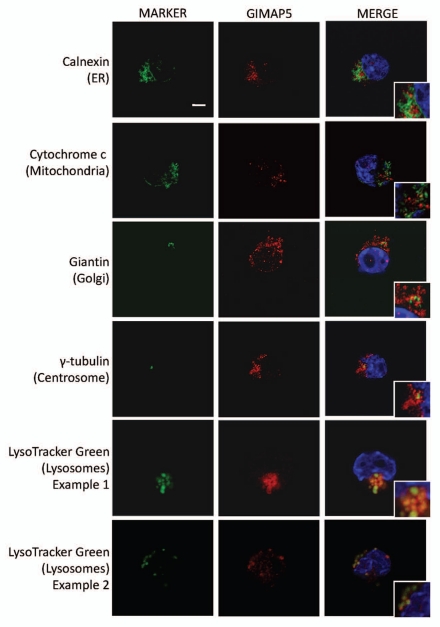
Figure 3.
Co-localization analysis in C1498 cells of mouse GIMAP5 with markers of different intracellular compartments. Representative images acquired by confocal microscopy of mouse C1498 cells stained as described in the Materials and Methods section. The intracellular compartments detected by the marker reagents (green) (see Table S2) are indicated to the left of the parts. Staining with anti-mGIMAP5 mAb MAC421 is in red. Cropped inserts display the key area of the parent image at ∼2-fold magnification. Nuclei (blue) were stained with DAPI. Scale bar = 5 µm.

