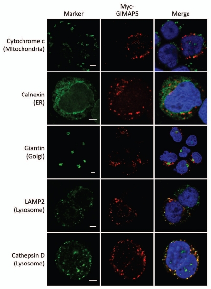
Figure 7.
Co-localization of human GIMAP5 with markers of intracellular compartments in Myc-huGIMAP5-TREx™ Jurkat cells. Representative images of the sub-cellular localization of myc-huGIMAP5 analysed by confocal microscopy are shown. Myc-huGIMAP5-TREx™ Jurkat cells (Fig. 6) were cultured in 1 µg/ml tetracycline for 24 hours before being processed for immunostaining. Localization of the myc-epitope tag (red) was compared with the indicated compartmental markers (green—indicated to the left of the parts) using antibodies as detailed in Table S2. Nuclei are stained blue (DAPI). Note that not all cells responded to tetracycline induction and hence some cells lack ‘red’ staining. Scale bars = 5 µm. An enlarged, 3D version of the image of cells co-stained for myc and cathepsin D is shown in Supplemental Figure 1.

