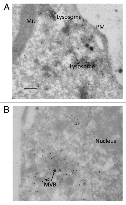
Figure 8.
Electron micrographs of anti-myc labelling of myc-hGIMAP5 in TREx™ Jurkat cells. Myc-huGIMAP5-TREx™ Jurkat cells were cultured in 1 g/ml tetracycline for 48 hours and then processed for electron microscopy as described in the Materials and Methods section. (A) Field selected to illustrate gold-labelling of electron-dense membrane vesicles recognisable as lysosomes (or related late endocytic vesicles); this field also shows the absence of label associated with mitochondria (Mit) and the plasma membrane (PM). (B) Field selected to illustrate association of gold label with structures recognisable as multivesicular bodies (MVB); this field also shows the absence of label associated with the nucleus. Scale bar = 200 nm.

