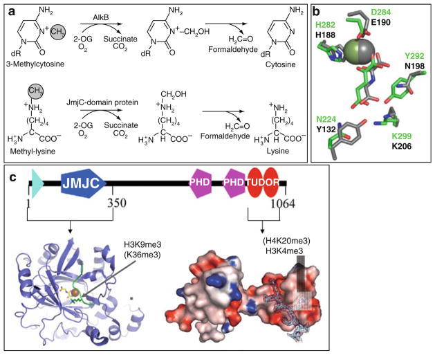
Fig. 4.
Demethylation by hydroxylation. (a) Mechanisms of demethylation of 3-methylcytosine by AlkB (top) and of methyl-lysine by Jumonji-domain proteins (bottom). (b) Coordinations of Fe2+ (sphere), α-ketoglutarate in JMJD2A (in gray), and KIAA1718 (in green). (c) Schematic representation of JMJD2A domain organization, including the structures of the N-terminal Jumonji (ribbons) [85] and the C-terminal double Tudor domain (surface representation) [87]

